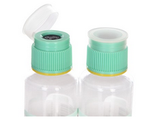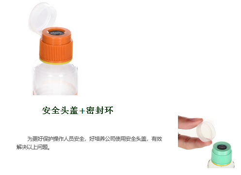
The "safety hood" effectively protects operators from acquired infections. During the blood collection process, the puncture point on the blood culture bottle stopper is exposed, and pathogenic bacteria may become infected through the respiratory tract or direct contact with the skin, eyes, or mucosal arch of the operator or laboratory personnel, posing a risk of laboratory acquired infections;
During the process of seed transfer, opening the lid to release air and extracting positive specimens can easily cause specimen leakage, posing a significant risk of infection to laboratory personnel;

The correct collection of clinical microbial specimens directly affects the results of microbial culture and identification. Reliable test results can guide clinical diagnosis and treatment, provide a basis for clinical scientific medication and successful infection control. It is the first and most important step in the correct and reasonable use of antibiotics, delaying bacterial resistance, reducing antibiotic abuse, and monitoring hospital infections. Therefore, all kinds of bacteriology specimens should be collected correctly. 1、 Collection of blood samples
1. Collection method
(1) Clean local skin with 75% alcohol.
(2) After the skin is dry, disinfect it with 2% -2.5% iodine from the center of the puncture point, with a range of no less than 5cm (diameter), and do not touch the disinfected skin with your fingers.
(3) After the skin iodine is dried (about 1 minute), blood is collected through puncture, with a blood collection volume of 5-10ml for adults and 1-5ml for infants and young children.
(4) Immediately after blood collection, inoculate the culture bottle next to the bed and quickly shake it gently. Mix thoroughly to prevent coagulation, but do not shake excessively to prevent hemolysis.
2. Precautions
(1) Suspected bacteremia should be collected as soon as possible. Blood collection with an increase in body temperature (38.5 ℃) can increase the positive rate, but it is also necessary to prevent delays in timing due to waiting.
(2) For those who have already used antibacterial drugs but cannot stop taking them, blood should also be collected before the next medication. Do not take blood samples from intravenous antibiotics, nor from venous catheters or arterial catheters.
(3) The ratio of culture medium to blood should be 10:1 to dilute antibiotics, antibodies, and other bactericidal substances in the blood; Some people advocate that for patients receiving antimicrobial treatment, the ratio of culture medium to blood collection can be 20:1 or greater.
(4) Recent studies have shown that replacing needles before injecting blood into blood culture media can actually lead to contamination.
(5) Blood samples should be collected at least twice per case, with an interval of 0.5-1 hours, in order to improve the positive rate and distinguish between infected and contaminated bacteria.
(6) For patients suspected of bacterial endocarditis and brucellosis, it is appropriate to take blood from the elbow or femoral artery. In addition to taking blood during the fever period, it is necessary to take blood for many times (3-4 times in 24 hours) and increase the amount of blood taken (10ml can be increased).
(7) After blood collection, it should be immediately sent for testing. If it is not possible to send for testing immediately, it can be placed at room temperature instead of in a refrigerator.
(8) If the clinical manifestation is similar to sepsis, but the blood culture is negative multiple times, it suggests considering the possibility of anaerobic and fungal infections.
2、 Collection of bone marrow
1. The collection method is to strictly disinfect the lesion site or the anterior (posterior) superior iliac spine, then extract 1ml of bone marrow and inject it into a blood culture bottle.
2. Precautions
(1) The bone marrow contains a large number of monocyte macrophage system cells, so bone marrow culture is superior to blood culture in the diagnosis of typhoid fever patients.
(2) Used for patients with suspected bacterial osteomyelitis.
3、 Collection of venous catheter specimens
1. Collection method
(1) Skin disinfection at the conventional catheter site.
(2) Sterile method: Extract the venous catheter from the patient's body, cut off the catheter tip 5cm, and place it in a sterile bottle.
(3) Immediately (within 15 minutes) submit for testing to avoid specimen drying.
2. Precautions
(1) Immediately inoculate the cut catheter into a blood plate to increase the positive rate.
(2) Repeatedly rinse the lumen of the catheter with broth, and conduct quantitative cultivation of the rinse solution.
(3) Someone placed the cut catheter in a meat broth enrichment solution or culture bottle, but this method cannot distinguish between catheter infected bacteria and a small amount of colonized bacteria.
(4) The Ministry of Health stipulated in the "Diagnostic Standards for Medical Infections" (Trial) (implemented on January 3, 2001) that the inoculation method for catheter tip culture should be 5cm from the catheter tip and rolled back and forth on the surface of the blood plate once. Bacteria ≥ 15cfu/plate are considered positive.
4、 Respiratory tract
1. Sputum specimen
(1) Collection method
① After waking up in the morning, rinse your mouth with cold boiled water multiple times to remove a large amount of miscellaneous bacteria in your mouth. Effort to cough up pus and phlegm from the deep lungs, and place it in a clean and dry container for examination.
② For those with minimal sputum volume, approximately 25ml of 3% -10% sodium chloride solution at 45 ℃ can be atomized. For young children with low expectoration, the trachea above the sternum can be gently compressed, and after expectoration, specimens can be collected using sterile cotton swabs.
③ Patients with weak cough or coma can use a sputum suction tube to attract sputum through the nasal cavity or oral cavity through the airway for examination.
④ Tracheotomy patients can penetrate deep into the ventilation holes to extract phlegm.
⑤ Bronchoscopy can be used to collect high concentration pathogenic bacteria directly at the focus.
⑥ Insert a sterile catheter into the tracheoscope through the nasal cavity, slowly inject 5ml of sterile distilled water, and remove the catheter. Collect the sputum coughed up by the patient within 3 hours and place it in a sterile container for examination.
⑦ Gastric sputum collection method. Tuberculosis patients without conscious symptoms. Sometimes phlegm can be mistakenly ingested into the stomach, so intragastric solution can be used for tuberculosis culture, and the positive result is about 10% higher than that of expectoration. This method starts in the morning on an empty stomach, and sterilized gastric tubes are fed into the stomach from the nasal cavity, and gastric juice is extracted using a 20ml syringe.
(2) Precautions
① The collection of sputum samples is based on morning sputum, when the patient has a large amount of sputum and a high bacterial content.
② Immediately submit the specimens for examination after collection. If they cannot be submitted in a timely manner, they can be temporarily stored in a 2-8 ℃ refrigerator. If inoculated 1-2 hours later, it will lose the aerobic bacteria and cause excessive growth of Gram negative bacteria.
③ Acceptable specimens: sputum; Bronchial lavage fluid, bronchial brush, bronchial biopsy; Lung aspirates and lung biopsies. Unacceptable specimen: saliva; Sputum left for 24 hours (excluding tuberculosis culture); Swab.
④ Quality evaluation of sputum specimens: (screened using a microscope) Under low magnification, examine squamous epithelial cells from at least 10 fields of view, focusing on areas with white blood cells. Qualified specimens have a ratio of white blood cells to squamous epithelial cells greater than 2:1.
2. Nasal swab
(1) Use a wet swab (physiological saline);
(2) Two nostrils need to be inserted;
(3) Go deep about 3.3cm and rotate 4-5 times;
(4) The above is used to diagnose nasal carriers during outbreaks of Staphylococcus aureus.
3. Pharyngeal swab, oral swab
(1) Collection method (take a specialized swab from the bacterial room)
① After the patient rinses his mouth with clean water, the examiner pulls his tongue out, and repeatedly wipes the posterior pharyngeal wall or the back side of uvula with a cotton swab for several times;
② When purulent tonsillitis or oral candidiasis occurs, directly wipe the lesion area with a cotton swab;
③ Insert into the transport medium and immediately send for testing to prevent drying.
(2) Precautions
① Cotton swabs should avoid touching the tongue, oral mucosa, and saliva;
② Do not rinse your mouth with disinfectant or come into contact with the lesion area a few hours before collecting specimens.
5、 Gastrointestinal tract
1. Fecal matter
(1) The collection method includes 2-3g of pus blood and mucus feces, and 1-2ml of flocculent material from liquid feces. Serve in sterile bottles or wax paper boxes for timely inspection.
(2) Precautions
① Fresh specimens should be collected, and outdated specimens affect the positive detection rate;
② Avoid mixing feces and urine;
③ If amoeba is to be separated, the specimen should be immediately sent for examination and attention should be paid to insulation;
④ Transporting the culture medium can increase the positive rate;
⑤ When separating Clostridium difficile, it is necessary to select unformed or watery stools;
⑥ Vibrio cholerae, Escherichia coli Ο The diarrhea caused by 157 infection is characterized by watery or bloody stools;
⑦ Samples are collected in the early stages of pathology or before treatment;
⑧ It is generally believed that after three days of using antibiotics, the specimen culture is negative for pathogenic bacteria.
2. In cases where feces cannot be obtained, an anal swab moistened with sterile glycerol water or sterile physiological saline can be inserted into the anus 4-5cm (2-3cm for young children), gently rotated and wiped to remove the rectal surface mucus, and then placed in a transport medium for examination.
6、 According to the Notice of the Ministry of Health on the Diagnostic Standards for Medical Infections (Trial), the clinical diagnosis of the urinary system is that the patient has urinary tract irritation symptoms such as frequent urination, urgency of urination, and pain in urination, or has tenderness in the lower abdomen, percussion pain in the renal region, with or without fever, and has one of the following conditions:; (2) Clinically diagnosed as a urinary tract infection, or recognized as a urinary tract infection due to effective antibacterial treatment.
1. Midrange urine
(1) Collection method
① Before sampling, women should rinse their external genitalia with soapy water or 0.1% potassium permanganate solution. They should urinate separately with their fingers and lips, discard their anterior urine, and do not stop urinating. Instead, 10ml of middle urine should be taken and stored in a sterile container.
② For males, clean the urethral opening with soapy water or disinfect the urethral opening with 0.1% iodophor solution. Dry the urethral opening with sterile gauze, fold the foreskin up, discard the anterior section of urine, and do not stop urination. Leave 10ml of the middle section of urine in a sterile container.
(2) Precautions
① Although catheterization can reduce pollution, repeated catheterization can cause retrograde infection, so in recent years, middle stage urine has been mostly used.
② It is best for female patients to take a bladder lithotomy position and have the nurse take the specimen to reduce pollution.
③ It is best to collect specimens for the first time in the morning.
④ After collecting specimens, if they cannot be submitted for testing within 1 hour, temporarily store them in a 4 ℃ refrigerator, but not for more than 8 hours. If the urine sample is left at room temperature for more than 2 hours, even if the bacterial count obtained by inoculation and culture is ≥ 104 cfu/ml and 103cfu/ml, it cannot be used as a diagnostic basis and should be re taken for testing.
⑤ When suspected of urethritis, the initial 3-4ml of urine can be collected in a sterile container. Even if there are a small number of bacteria in the urine, if they are repeatedly tested for the same bacteria, they should still be considered as pathogenic bacteria.
⑥ In most cases, flushing the external genitalia alone is not enough for children, so collecting specimens is more difficult. If there is a significant increase in bacteria in the urine, it can be highly suspected of being a urinary tract infection, and if it is sterile, it can be denied.
⑦ If the bacterial culture results in two or more types of bacteria, it is necessary to consider the possibility of contamination, and it is recommended to keep a new sample for testing.
⑧ Preservatives and disinfectants should not be added to urine, otherwise it will affect the positive detection rate.
2. Bladder puncture collection method is very difficult to avoid pollution. When the cultivation results are not consistent with the condition, or when urine anaerobic bacteria cultivation, or when it is difficult to collect mid stage urine in infants and young children, suprapubic puncture can be used to collect urine from the bladder.
3. The collection method of indwelling catheter is to pull out the urine collection bag with closed drainage, discard the urine in the front of the catheter, and leave 10ml of uncontaminated urine in the bladder for inspection. Do not leave specimens from the lower end of the urine collection bag.
7、 Cerebrospinal fluid
1. Collection method: Clinicians collect 3-5 ml of cerebrospinal fluid (1 ml of bacteria, 2 ml of fungi, 2 ml of acidfast bacteria, and 1 ml of viruses) in sterile tubes or vials and immediately send them for testing.
2. Precautions
(1) Samples were immediately sent for examination after collection. Due to the rapid autolysis of Neisseria meningitidis after isolation, Streptococcus pneumoniae and Haemophilus influenzae were also prone to death;
(2) When suspected to be infected with the aforementioned bacteria, attention should be paid to insulation and should not be placed in a refrigerator or stored at low temperatures.
8、 Bile
1. Duodenal drainage method Under aseptic operation, when swallowing the duodenal duct to the duodenal papilla (65-70cm from the teeth), collect solution A (from the common bile duct, orange or golden), then inject 40ml of 25% magnesium sulfate solution, after 1-2min, take solution B (from the gallbladder, brown yellow green), and then collect solution C (from the liver bile duct, lemon color). It is generally believed that solution B is of great significance for bacterial culture.
2. During gallbladder puncture cholecystography, bile can be taken simultaneously.
3. The surgical method is to directly puncture the common bile duct and gallbladder to collect bile. The samples collected above should be immediately sent for examination, otherwise they should be placed in a 4 ℃ refrigerator. Care should be taken when collecting to avoid bacterial contamination from saliva and duodenal fluid.
9、 The puncture fluid sample includes pleural fluid, ascites, pericardial fluid, joint fluid, and synovial fluid.
1. The collection method is performed by a clinical physician for puncture extraction. Chest and ascites can be collected in 5-10ml, pericardial fluid and joint fluid in 2-5ml, placed in sterile test tubes or vials containing anticoagulants, thoroughly mixed, and immediately sent for examination. Anticoagulant 10% EDTA disodium salt, sample to anticoagulant ratio 10:1.
2. Precautions
Specimen collection should be conducted before the patient takes medication or 1-2 days after discontinuing medication;
(2) If it cannot be immediately sent for inspection, it can be stored in a 4 ℃ refrigerator;
(3) Joint fluid from patients suspected of gonococcal arthritis should be immediately collected and sent for examination, or it is better to have it cultured by the bed;
(4) Do not use a cotton swab to dip the sample for testing.
10、 Abscesses and lesion secretions
1. Wound
(1) Surface wound swab collection method and precautions
① Limited to skin and subcutaneous tissue, including surgical incisions, bedsore ulcers, neonatal omphalitis, and infant abscesses.
② Wipe off the surface exudate of the lesion with sterile physiological saline or 75% alcohol;
③ Use a sterile cotton swab to wipe off and remove deeper pus secretions or tissues for examination;
④ When collecting specimens of bedsore ulcers, purulent secretions from the edge of the bedsore should be collected, and the swab should be placed in the transport medium for immediate examination. Do not collect exudate from the wound surface.
⑵ Collection methods and precautions for wounds, abscesses, sinuses, and gangrene tissues
① After disinfecting the skin with iodine and alcohol for closed abscess, use a sterile syringe to puncture and extract all pus for examination. If it is suspected to be an anaerobic infection, remove the air from the needle tube and insert the needle into a sterile rubber stopper for examination;
② If there is pus and exudate at the wound, use a sterile cotton swab to go deep into various sinuses to wipe, scrape with a knife, puncture and aspirate, or perform surgical resection to obtain deep wound samples;
③ When there is bleeding from trauma, specimens should not be collected within 2 hours of medication application or 12 hours of burns, and there is very little chance of obtaining positive results at this time.
④ Pay attention to observing the characteristics, color, and odor of pus and secretions when collecting specimens. It can provide a basis for cultivation and identification.
⑶ Burn wound surface
① Burn wounds require purulent secretions, and the eschar quickly separates and turns brown, black, or violet colored, with edema at the edges of the burn. The patient has a fever>38 ℃ or a low body temperature<36 ℃, accompanied by hypotension.
② Rinse the wound surface with sterile physiological saline.
③ Due to different wound sites and different types of bacteria, it is necessary to use sterilized cotton swabs to collect inflammatory areas from multiple sites for examination.
④ Collect the edges or drainage fluid of unhealthy areas with blackening or degeneration under the scab for quantitative culture and tissue biopsy.
2. Anaerobic wound swab
(1) Anaerobic cotton or glass rod touches deep pus;
(2) A syringe can be used for suction to remove air from the needle tube and insert the needle into a sterile rubber plug for inspection;
(3) Immediately submit for inspection, and maintain oxygen free conditions during the inspection process;
There are sputum, throat swabs, nasopharyngeal swabs, gingival swabs, rectal, vaginal, and cervical swabs, as well as effluent from ileostomy and colonostomy, as well as contents of the stomach, small intestine, and large intestine that are not suitable for anaerobic bacterial cultivation.
3. Urethral discharge swab
(1) Men use sterile gauze to wipe and clean the urethral opening, and then take purulent secretions that overflow from the urethral opening;
⑵ When collecting prostatic fluid, first flush the urethra and bladder, then massage the prostate with fingers from the anus to promote the overflow of prostatic fluid;
(3) Female patients should first carefully wipe or clean the urethral opening with sterile gauze or cotton swab, and then compress the urethra from the vagina, causing secretion to overflow;
(4) Take cervical secretions, first use an endoscope to dilate the vagina, and then use a sterilized cotton swab to collect the secretions from the cervix, trying to avoid contamination by normal bacterial communities near the vagina and cervix.
4. Cellulitis
(1) Disinfect the skin with sterile physiological saline or alcohol;
(2) Use a fine needle to puncture and attract the most prominent area of inflammation;
⑶ Draw a small amount of sterilized physiological saline into the needle tube;
Inject into sterile test tubes for testing.
5. Tissue specimen
Micro samples taken from living tissues can be directly inoculated into the culture medium;
(2) The superficial tissue can be directly wiped with a cotton swab or scraped off with a small knife;
(3) The surface of larger tissue blocks can be cauterized or immersed in boiling water for 5-10 seconds, and then cut open with sterile scissors to remove the pus;
Deep tissue specimens can be obtained and submitted for examination through skin puncture or surgical incision;
(5) Tissue specimens cannot be fixed with formalin; (6) Sometimes the innermost dressings stained with pus can also be placed in sterile plates for examination.
11、 Eye specimen collection
(1) For general bacteria, use sterile cotton swabs to collect eye secretions, especially purulent secretions;
(2) Chlamydia trachomatis, wipe off first


